
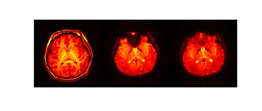
One way to "recover" the signal is by using an RF excitation pulse that is designed to "precompensate" the phase of the spins in the slice for the dephasing effects of susceptibility. Chun-yu Yip, an EE:Systems graduate student who is jointly mentored by Jeff Fessler and Doug Noll, has developed an iterative method for designing RF pulses that are "tailored" to the specific properties of each person scanned. (Work accepted to appear in Magnetic Resonance in Medicine in 2006.) The right hand image below shows a T2*-weighted image that was collected using this special RF design approach. There is substantial signal recovery in the mid frontal brain regions above the sinus cavity.
This type of sophisticated signal processing
will enable neuroscientists
to study cognitive processes
in brain regions
that were previously
very diffult to see
with fMRI.